

With thousands of mares foaling every year, it is common for veterinarians to be called to see a mare that has not lost her placental membranes after foaling. Most of the time, retained foetal membranes (RFMs) can be dealt with easily and without complications, but they can result in disastrous consequences if not treated in a timely fashion.

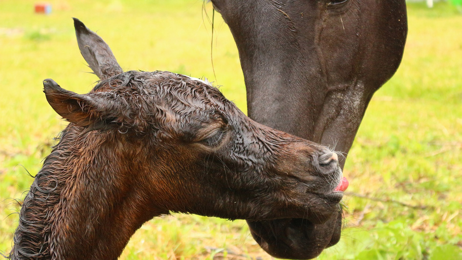
Foetal membranes should be passed shortly after the delivery of the foal. © Maxine Brain.
The placenta is formed by the foetus and starts its development around 35 days (34-37) when cells from the developing foetal unit migrate into the endometrium (uterine lining) of the mare to form what are called endometrial cups. The placenta continues to develop and expand throughout the uterus until it is physically in contact with the entire endometrium. The foal develops predominantly in one side of the uterus or the other, depending on whether it implanted at the junction between the horn and the body of the uterus on the right or the left side.
The placenta itself consists of two tissues – the chorion and the allantois – that fuse together, with the allantois being the surface on the foetal side, and the chorion, the surface on the maternal side. It is the chorionic layer of the placenta that has the bright red velvet appearance due to the small, finger-like projections of blood vessels. These placental blood vessels interlace with the endometrium vessels, forming a tight bond that persists until the foal is born.
It is this interaction of the placental structure with the endometrium that provides oxygen, nutritional support, and waste removal to the foal during its term in the uterus. The placenta/uterus apposition forms a unique functional unit that allows oxygen and nutrients to pass from the maternal blood circulation into the blood vessels of the placenta, and into the foal via the umbilical cord, and the return of carbon dioxide and waste products from the foal back through the placenta and into the mare’s blood system to then be expelled from her body.
As the mare goes through the second stage of labour and the foal is delivered, the umbilical cord ruptures causing a drop in blood pressure, which, together with the rhythmic contractions of the uterine muscle, brings about the final stage of labour, the release of the placenta and its expulsion from the uterus. In the typical foaling scenario, the placenta is expelled within three hours of the foal being delivered, and more commonly within one hour. The placenta is usually expelled inside out, so that the white side of the allantois is the side we see, with the red chorion on the inside.
VETERINARY ASSISTANCE
Foetal membranes that are not released within three hours are generally referred to as delayed foetal membranes and should alert the owner that retention of the membranes is a strong possibility and plans for veterinary advice or intervention should be sought. Membranes that have not been expelled by 6-8 hours are referred to as an RFM and require veterinary assistance as soon as possible. Typically, it is the non-pregnant horn of the placenta that is the last part of the membranes to detach from the uterus.
Draught mares have a reported increased incidence of RFMs, as do pregnancies that have a prolonged gestational length or compromised delivery such as a dystocia or caesarean. RFMs can also occur following an abortion and sometimes this is the only evidence that alerts the owner to the loss of a pregnancy.
The initial treatment for RFMs is the administration of oxytocin, a hormone that causes uterine contractions. This can be done when membranes have not been passed within an hour of birth and is maybe all that is required. Oxytocin can be administered several times at 1–2-hour intervals to encourage the uterus to contract and the membranes to be expelled.
If the membranes are not expelled using oxytocin alone, then it is common practice for the veterinarian to do a uterine lavage or flush to encourage the membranes to be released. This can be done several ways: one is to infuse fluids, often isotonic fluids (such as Hartmann’s Solution) directly into the uterus to cause it to expand and induce muscle contractions. This fluid is then allowed to be passed out through the vulval lips, taking with it old blood clots and foetal fluid remnants that may also be sitting in the uterus encouraging the proliferation of bacterial organisms. Some veterinarians add either an antiseptic or antibiotics to the fluid to assist in reducing the risk of an infection occurring.
The second way is to fill the inside of the placenta with warm water and to hold the water in there by either tying the open end of the placenta, or just using a tight fist around the opening to stop the fluid escaping. This causes the uterus and the placenta to expand and release but relies on the placenta being intact and not torn. Water is sufficient to use on its own as the fluid is retained within the placenta and does not contact the uterus and is passed out once the pressure around the opening is released. This is not suitable to use if the placenta is torn, as the water will escape into the uterus, which is not desirable.

Membranes that are still attached several hours after the foal has been born. © Maxine Brain.
DEBATABLE PROCEDURE
Manual manipulation to remove the RFM is a debatable procedure, and if done it needs to be performed by an experienced veterinarian. Some veterinarians recommend the membranes are left in situ and the mare flushed daily until the membranes are released on their own. They believe the risk of damaging the uterus or tearing the membranes is too high if manual manipulation is performed. Personally, I have removed many RFMs by gentle manipulation and seldom incurred a problem, however, it is a lengthy and slow procedure and cannot be rushed. If performed by an inexperienced person, there is a high risk that the placenta will tear, leaving behind remnants of membrane that can cause a life-threatening infection in the uterus, or worse, induce a uterine prolapse that can kill the mare if not replaced swiftly. Unfortunately, I know of cases where the owner has tried to unsuccessfully remove the RFM and the end result was the death of the mare.
For veterinarians that do not prefer to manually remove the membranes, daily broad-spectrum antibiotics and uterine flushing is required as the risk of infection and other life-threatening conditions increases with every day the membranes remain attached.
Whilst it is good to tie the placenta up to stop it dragging or being caught on something and tearing, the practice of tying a heavy object to the placenta that is exposed and hanging out from the vulval lips is not recommended. Suspending a heavy weight can have the same consequences as having an inexperienced person pulling on the membranes, causing tearing or uterine prolapse if the membranes are firmly attached.
Once the placenta has been successfully removed from the uterus, the membranes should be assessed to ensure that all the membranes are out and that there are no tears or missing pieces that can cause a serious infection. Generally, the uterus should be flushed at least once again after the placenta is removed to encourage the evacuation of uterine fluids and bacteria that have accumulated. Most mares benefit from daily oxytocin injections in the days following an RFM to encourage the involution of the uterus and the expulsion of fluid. Antibiotics are not required unless there are membrane fragments remaining, or the mare has developed a systemic infection from a damaged endometrium. Anti-inflammatories such as Finadyne are often given to provide pain relief and reduce the risk of the secondary complications that may be seen with RFM.

This is a RFM that was removed. The white surface is the foetal side and the red surface is the side that attaches to the uterus. © Maxine Brain.
“The message for all owners is to check
their mares shortly after foaling.”
SEVERAL COMPLICATIONS
There are several complications that can develop if an RFM is not dealt with quickly and effectively and these include systemic illness in the mare, future infertility issues and laminitis. The first, systemic illness, essentially means the mare becomes ill from either bacteria or toxins being absorbed into the mare’s bloodstream from the RFM and causing an infection.
Infertility can result from damage caused to the endometrium due to infection in the uterus or from physical damage caused when the RFM is forcibly removed. This can take the form of the mare failing to conceive, conceiving an embryo but losing it early in the pregnancy, abortion of a foal late in the pregnancy or the mare delivering a small, weak foal that can die if not treated intensely.
The third recognised complication is laminitis caused by the absorption of endotoxins from the uterus. This can be devastating as, unlike many cases of laminitis seen with hormone associated laminitis (Cushing’s and Equine Metabolic Syndrome), where the laminitis can be mild and amenable to treatment, endotoxic induced laminitis is often severe and rapid in onset, causing profound lameness and stress. Any mare that has had an RFM should have their feet checked for signs of heat or increased digital pulses to ensure early signs are detected and appropriate treatment plans put in place.
The message for all owners is to check their mares shortly after foaling, taking note that there is nothing unusual hanging from the vulval lips. If they see membranes attached, contact their veterinarian promptly and make plans to ensure their mare receives the appropriate attention at the right time, so her health and the wellbeing of her foal are not compromised unnecessarily. EQ
YOU MIGHT ALSO LIKE TO READ:
Preparing for Laminitis – Equestrian Life, September 2022
Working Together for Best Outcomes – Equestrian Life, August 2022
What Constitutes an Emergency – Equestrian Life, July 2022
Peri-Tarsal Cellulitis Calls for Quick Action – Equestrian Life, June 2022
Sinusitis: Not To Be Sneezed At – Equestrian Life, May 2022
Japanese Encephalitis: No Cause For Alarm – Equestrian Life, April 2022
Hernia Learning Curve – Equestrian Life, March 2022
Osteochondromas: Benign But Irritating – Equestrian Life, February 2022
Don’t Forget the Water – Equestrian Life, January 2022
Understanding Anaesthesia – Equestrian Life, December 2021
A Quick Guide to Castration – Equestrian Life, November 2021
Caring for Mammary Glands – Equestrian Life, October 2021
Sepsis In Foals – Equestrian Life, September 2021
Understanding Tendon Sheath Inflammation – Equestrian Life, August 2021
The Mystery of Equine Shivers – Equestrian Life, July 2021
Heads up for the Big Chill – Equestrian Life, June 2021
The Ridden Horse Pain Ethogram – Equestrian Life, May 2021
The Benefits of Genetic Testing – Equestrian Life, April 2021
Heavy Metal Toxicities – Equestrian Life, March 2021
Euthanasia, the Toughest Decision – Equestrian Life, February 2021
How to Beat Heat Stress – Equestrian Life, January 2021
Medicinal Cannabis for Horses – Equestrian Life, December 2020
Foal Diarrhoea Part 2: Infectious Diarrhoea – Equestrian Life, November 2020
Foal Diarrhoea (Don’t Panic!) – Equestrian Life, October 2020
Urticaria Calls For Detective Work – Equestrian Life, September 2020
Winter’s Scourge, The Foot Abscess – Equestrian Life, August 2020
Core Strengthening & Balance Exercises – Equestrian Life, July 2020
The Principles of Rehabilitation – Equestrian Life, June 2020
When is Old, Too Old? – Equestrian Life, May 2020
Related articles

Australian Lipizzaner enthusiasts are leading the world with a global database for the famous breed and attracting international entrants for the first Lipizzaner Global High Point competition.
Australia’s eventers gave it their best shot but found the competition unrelenting, the courses daunting and their competitors’ horses “beasts that you had to see to believe”!